Using high-density surface electromyogram to identify innervation zone in the external, anal sphincter muscle
Innervation zones, where end-plates distribute within striated muscles, can be identified with arrays of electrodes. For this to be possible, electrodes and muscle fibres must reside in parallel planes. Innervation zones are identified where two, consecutive action potentials with opposed polarities can be found. In each direction from this location, action potentials are detected with progressively larger delays, as they propagate along the muscle fibres.
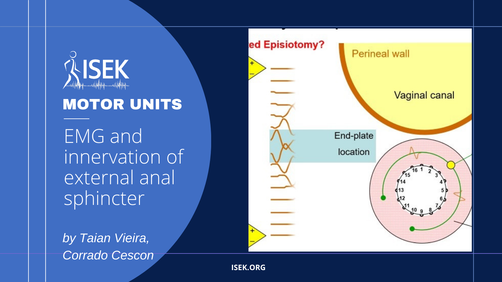
The material shared here illustrates how innervation zones can be identified for a particular, striated muscle—the external, anal sphincter (EAS). Given the EAS fibres arrange concentrically to the anal canal, defining their beginning and end (green and blue circles in slide 1) is not possible. However, with electrodes disposed on a cylindric support, action potentials propagating along the EAS fibres can be detected, so allowing for the identification of innervation zones (slide 3). This knowledge is expected to help guiding the practice of episiotomy, ensuring minimal, nerve damage (slide 2).
About the Authors

Corrado Cescon
University of Applied Science of Southerns Switzerland
Corrado Cescon is a senior researcher at the Rehabilitation Research Laboratory (2rLab) of the University of Applied Science of Southerns Switzerland (SUPSI). His research interests are EMG signal processing, image processing and motion capture analysis.

Taian Vieira
Politecnico di Torino
Taian Vieira is with the Laboratory for Engineering of the Neuromuscular System (LISiN), Politecnico di Torino, Italy. His research activities focus on the extraction of information of valid, applied interest from surface electromyograms.


