EMG descriptors

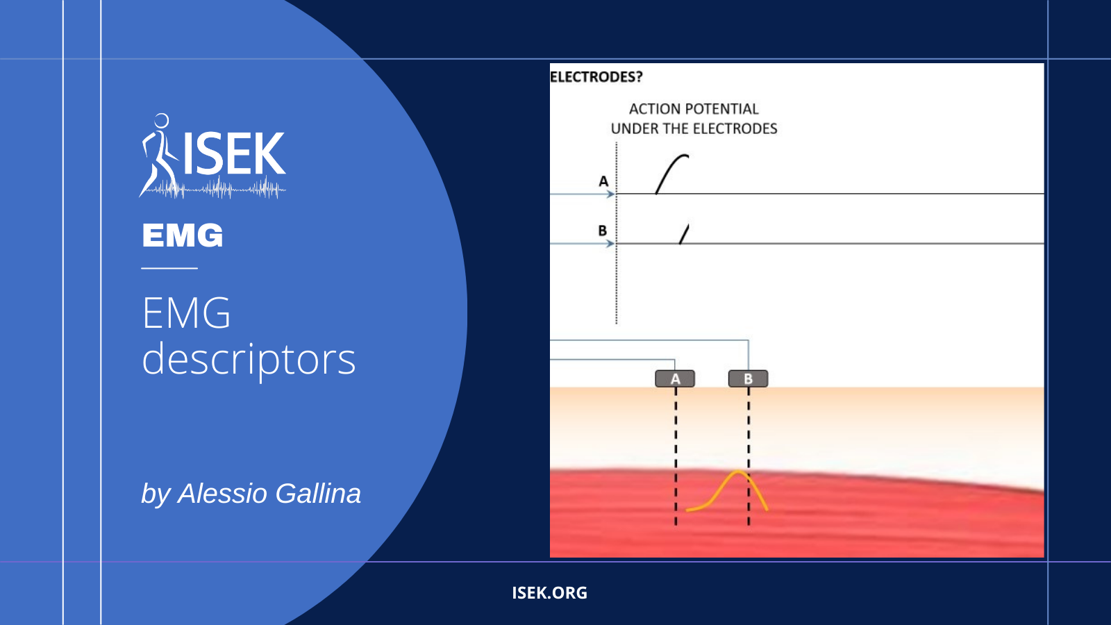
This powerpoint presentation contains 11 animations. Slide 1 illustrates the generation of the muscle fiber action potential and how it contributes to the generation of electromyographic signals. Slides 2-3 illustrate why two electrodes are commonly used to record electromyographic signals, and specifically why the bipolar detection helps reducing the electrical interference. Slides 4-11 illustrate how changes in motor unit firing rate, number of recruited motor units, motor unit depth and muscle fiber conduction velocity are reflected in EMG amplitude estimates.

DOWNLOAD POWERPOINT

About the Author

Alessio Gallina

Alessio Gallina

University of Birmingham

Alessio Gallina has prepared this material while employed at the Laboratory for Engineering of the Neuromuscular System, Politecnico di Torino, Italy. He is currently employed as Assistant Professor in the School of Sport, Exercise science and Rehabilitation, University of Birmingham, United Kingdom.

https://scholar.google.it/citations?user=58Y8OnYAAAAJ&hl=en

COPYRIGHT

(c) 2023 by the author. Except as otherwise noted, the ISEK Teaching Repository, including its text and figures, is licensed under a Creative Commons Attribution-ShareAlike 4.0 International License. To view a copy of this license, visit https://creativecommons.org/licenses/by-sa/4.0/legalcode.

You May Also Like…

Muscle force regulation

Muscle force regulation

Passive and active mechanisms determine the net, muscle force.  The latter is associated with the number of muscle fibers excited, and thus motor units recruited, and the rate at which muscle fibers are excited.  The first three slides provide an approximate...

HD-EMG and action potential propagation

HD-EMG and action potential propagation

The muscle fibre conduction velocity (CV) is the speed with which action potentials travel along the muscle fibres. Owing to its sensitivity to events of applied interest (e.g. muscle fatigue), CV earns relevance for the study of the neuromuscular system. The...

EMG and innervation of external anal sphincter

EMG and innervation of external anal sphincter

Innervation zones, where end-plates distribute within striated muscles, can be identified with arrays of electrodes.  For this to be possible, electrodes and muscle fibres must reside in parallel planes.  Innervation zones are identified where two, consecutive action...